Even Scanning Electron Microscope manufacturers have marketers…

I got an unsolicited email asking for help boosting search-engine rank. Me! Complete with the particular phrase they wanted linked and everything. Normally, I’d be inclined to sneer and make mocking comments about marketers…except in this case it’s actually for something completely relevant…and kind of nifty, so I’m going to do it…

A tiny Scanning Electron Microscope manufactured by ASPEX A company called ASPEX makes what they’re calling an “Affordable Desktop SEM”. Not having a gigantic corporate budget, government grants, or wealthy patron/matron backing me I’m having trouble thinking of ANY Scanning Electron Microscope (there’s that phrase and link…) as being “affordable”, but I’ve got to admit I want one now.

That’s not all, though – ASPEX (no, I don’t know why they capitalize the whole name) has a mind-blowingly cool offer going on right now as part of their promotional campaign. “Send Us Your Sample” – just like the name suggests, they’re soliciting samples to image and post. I know I’m planning to go for it. It’d be nice to have both old-school light-microscopy images AND Electron microscopy imagery of Fred (my sourdough culture in progress) to post.

I’m not sure what kind of magnification they can offer – The submission form is pretty simple but doesn’t say – but I’d be really interested to know if I’ve got any bacteriophages in my sourdough slowing down the lactic acid bacteria growth…

I would like to add that I’m getting no particular compensation for this post, but if anyone from ASPEX corporation wants to upgrade me to “paid shill”, I wouldn’t say “no” to an SEM of my own…

No? Dang.

Anyway, for anyone who hasn’t given up on my blog despite the slow updates lately, thanks. There will be more photos, along with other impending microbiological nerdity. And food – sadly, I have not managed to get time to actually make the first of the new pie that I’ve invented in time for Pi Day (“3.14″…March 14…”Pi day”. Insert joke about doing something at 1:59am and laugh track sound here…), but “pigsfly pie” will actually exist soon. Honestly.

Microscopy Preview

Intentional food microbiology:
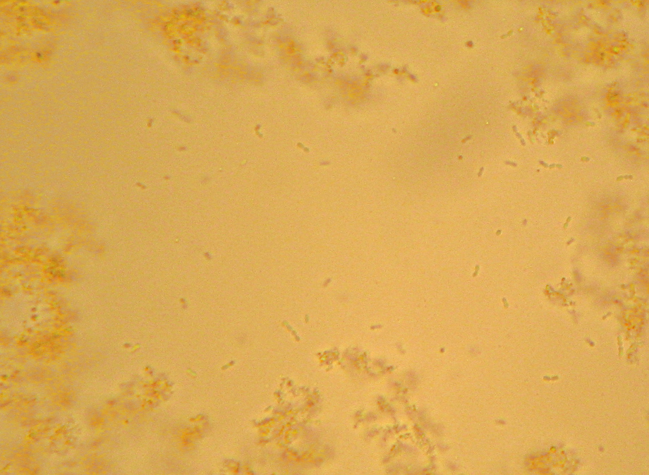
Various yogurt bacteria floating amid milk protein and fat globules

UNintentional food microbiology:
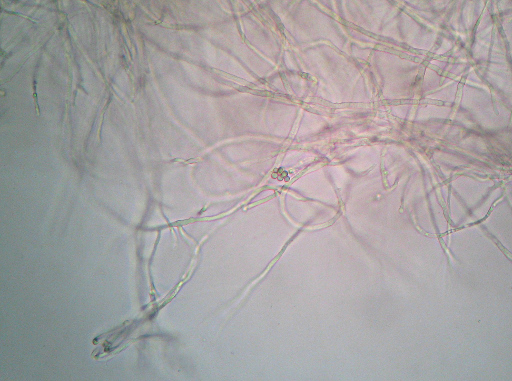
Wet-mount photomicrograph showing mold mycelia and a mass that kind of looks like large spore cases or something

I still don’t feel like I got nearly enough productive stuff done this weekend, but I did manage to do a bit of microscopy – plus demonstrating to myself that I still remember how to do a “Gram stain”. Real Post with explanation and more pictures to follow Real Soon Now…

Ewwwwwwwwwwwwww……

They say “When life gives you lemons, make lemonade”. What if life gives you snot instead?

Someone's slime-covered handNow, see, this is what happens when I’m too poor to buy nice distracting new toys for myself. (No, not the hand in the picture – that’s not mine, it’s just there for illustration.)

I found a pot that I’d rinsed well but then left filled with water in the sink to soak, to help remove the last of the rice bits stuck to it. It hadn’t gotten stinky or fuzzy or anything, but it had gone…viscous. Like a light sewing-machine oil. Naturally, I took appropriate action to deal with it.

I fed it.

Glucose (“dextrose”), to be precise. It’s since been dumped into an old glass jar and the original pot thoroughly scrubbed with hot soapy water. At this point (a day later) the slime is closer to the viscosity of vegetable oil now. And I fed it again.

I wonder what it is? I mean, obviously it’s bacteria-snot, but what kind? I suppose if I had some iodine I could check to see if it’s a polysaccharide (evidently this test works on polysaccharides besides starch). If only I had a microscope, I could at least get some basic hints as to what’s producing the slime. Maybe I can maintain a culture and figure it out later, if I can ever afford a real microscope. Perhaps I could even attempt a strain-improvement program to increase the production rate…

Uh…I did mention I was a nerd, right? Okay then.

I wonder if anyone at work has a bacteriological microscope setup that I could use?…

“Ueber die isolirte Faerbung der Schizomyceten in Schnitt- und Trockenpraeparaten”

The Giant’s Shoulders blog carnival is coming up in two days, and I just realized I still haven’t gotten a post up for it yet. So, here it is.

I put up some quick reviews of several classic microbiology-methods papers for the previous edition of this blog carnival, but didn’t actually get around to putting up the one for what is almost certainly the most well-known microbiology technique: “The Gram Stain”. So, this post is about it:

Gram HC: “Ueber die isolirte Faerbung der Schizomyceten in Schnitt- und Trockenpraeparaten”; Fortschritte der Medicin; 1884; vol 2, pp 185-189

That’s “Regarding the Isolational(?) Coloring of Schizomycetes in Cut- [i.e. tissue sections] and Dried Preparations” in “Medical Progress”. The translation hosted by the American Society for Microbiology uses the word “Differential” where I’ve put “Isolational” – which is probably not quite right either but it’ll have to do for now – but I’ll get to that in a moment.

If you’ve ever been exposed to microbiology labwork before, you’ve almost certainly done or at least watched a procedure referred to as a “Gram stain”. In brief, you smear your sample with bacteria on a glass slide and bake it on, then you dump some purple stuff on it, them some brown stuff, then you rinse it briefly with alcohol, then you dump on some pink stuff, and then rinse it in water and look at it under a microscope. Bacteria that stay the original dark purple-blue color of the original purple/brown stuff are considered “Gram Positive”, and those that don’t instead appear the pink color of the last stain, and are considered “Gram Negative”. Many textbook authors and microbiology instructors will breathlessly proclaim that the Gram Stain reveals two “fundamental” categories of bacteria, but I’ll spare you my rant about that.

Properly speaking, this isn’t actually Gram’s stain, as described in his original paper. The modern variations that we’re all taught in microbiology class were developed later, and I believe they are nowadays based mainly on Victor Burke’s 1922 paper on the subject[1].

Regarding the title of the paper: “schizomycete” is what they used to call most kinds of bacteria. “Mycete” meaning “fungus”, as bacteria were assumed to be “plants without chlorophyll” just like molds and mushrooms, and “Schizo-” meaning “split in two”, since bacteria reproduce by splitting into two cells rather than by producing spores like “other” fungi. I say “most” because things like cyanobacteria (“blue-green algae”) or Green Sulfur Bacteria would have been referred to as “Schizophyta” (“fission-plants”). What Gram was originally trying to do wasn’t to differentiate one kind of bacteria from another, either, but to make it easy to tell bacteria from from the nuclei of cells in bacteria-infected tissue.

For that matter, Gram was really metaphorically standing on the shoulders of Koch and Erhlich, as he was building on their technique for staining “tubercle bacteria” – that is, tuberculosis-causing members of the genus Mycobacterium. Gram mentions that you need to stain this type of bacteria for the “usual” 12-24 hours to make this work, incidentally, as opposed to a few minutes for other “schizomycetes”. This suggests that you are expected to have some idea of what you’re going to find before you use the stain, as opposed to the modern implementation which is supposed to tell you something about what kind of bacteria you’re finding.

Still, Gram does report that some bacteria take the stain and some don’t, giving us a preview of the “differential” character of the modern version. He specifically notes typhoid and some causes of bronchial pneumonia fail to hold the stain. Given that Typhoid Fever is caused by a strain of the “Gram-negative” butt-bacter Salmonella enterica, and there are a number of “Gram negative” bacteria as well as “Gram positive” that can cause pneumonia, this makes sense. He also does mention the use of Bismarck Brown R a.k.a. Vesuvine as a counterstain in order to make the nuclei of the infected cells brown in contrast to the dark blue of the infectious bacteria in the tissue.

For much of the century-and-a-quarter since Gram’s publication, the question of why the Gram stain works was thoroughly investigated, and even today I occasionally hear or read assertions to the effect that the Gram Stain isn’t well understood. I disagree with this just as I think its importance to bacterial identification is grossly overblown, and if you want to know why, I have a previous post all about why the Gram stain works and how we know. You may or may not also be interested in an older post regarding whether or not “acid-fast” bacteria like the ones that cause tuberculosis (which don’t stain at all when using the modern version of the Gram stain) are “Gram Positive” or not. As always, if you spot any errors or have any questions, please let me know…

[1] Burke V: “Notes on the Gram Stain with Description of a New Method.” J Bacteriol. 1922 Mar;7(2):159-82.

“A simplified method of staining endospores”

One more for the “classic papers” challenge:

Schaeffer AB, Fulton MD: “A Simplified Method of Staining Endospores”; 1933; Science; 77; pg 194

If you take a microbiology lab, this is the endospore staining technique (or “technic” as they used to spell it) that you’ll practice. This is a nice, simple, one-page paper. Alice B. Schaeffer and co-author Mac Donald Fulton describe a few of the other variations on endospore staining techniques, then describe how they’ve further simplified what they felt was previously the simplest one, described by a Mr. Wirtz in 1908.

“Endospores” are a sort of “escape pod” for certain specific kinds of bacteria. Unlike spores formed by yeasts and molds, these are not reproductive – each bacterium only produces one thick-coated spore, into which it shoves it’s genetic material and a few vital enzymes to get itself going again later when the spore finds itself in favorable conditions.

Since only a few kinds of bacteria produce these endospores, if you see endospores in your unknown bacterial culture it goes a long way towards helping to identify the bacterial species, so having a simple method for staining your bacteria so that endospores are obvious under a microscope is helpful. (Of course, these days most of us would rather just get a 16s rDNA sequence with PCR, but never mind that for now…)

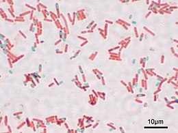
Endospore stain under a microscope (via Wikipedia)Evidently, Wirtz’s original method involved using Osmium Tetraoxide (“osmic acid”) to stick the bacteria to the slide before staining. Not only is that stuff poisonous, it’s also expensive. I found a site selling sealed glass ampoules containing 1 gram each of this stuff for $35.00 each. Schaeffer and Fulton’s method does away with this in favor of much cheaper and easier heat-fixing (just as is done with the Gram stain and others). They use the dye “Malachite Green” for the initial stain, and steam-heat the dye-covered bacterial slide a few times to sort of “cook” the dye into the thick-walled endospores if they are there. Rinsing then washes the dye out of everything but the endospores, and a light red dye (safranin) is added as a counterstain. The end result is that under the microscope you’ll see light-red bacteria. If any of them form endospores, you’ll be able to see them as smaller green dots – sometimes still bulging inside of bacterial cells, sometimes floating around freely having escaped from the now-empty bacterial cell.

The “Schaeffer-Fulton Endospore Stain” is pretty easy to do, though the occasionally messy steambath part can be annoying. The method is pretty resistant to errors, so it’s not too hard to get good results even if you’ve never done it before.

Incidentally, you can buy Malachite Green at many pet stores – it’s still used as a treatment for “ick” (Ichthyophthirius infestation) in tropical fish.

Hmmm…still a couple of hours before it’s not longer May – Perhaps I can throw in one last post before time’s up…

New toy: “Twitter”

Wow – Celestron takes 8 business days to get me a terse one-sentence answer. BigC responds in one. Impressive. Apparently their technical people are all at trade-shows at the moment so my bigger question will have to wait until they get back, but they were at least able to answer my question about their “tabletop” digital microscopes magnification (answer: the “600x” really is optical magnification, not digital.)

Another digital microscopy WANT/DO NOT WANT post to follow when I get the followup reply. Meanwhile, after hearing about it on the This Week in Tech podcast for a while, I finally talked myself into signing up to play with the coincidentally named Twitter system.

Twitter logoIt sounds like a really stupid idea – “Oh, goodie, now I can broadcast ‘text messages’ no more than 140 characters long about trivial events in my life to the whole world! Whoopee!” “Wow! I can find out when random strangers are drinking coffee AS IT HAPPENS!” Thrills! Excitement! Adventure!…

On the other hand, having the messaging system watch for particular words might be a handy way of monitoring current events. Plus, there seems to be a lot of potential for fun, off-the-wall uses, even if many of them are kind of silly.

It DOES seem like kind of an ideal context to play with that “geostrings” concept I’ve been toying with. A terse, easily-machine-parsed format for geotag data that can fit into a “twitter” post and still leave room for a sentence or two to go with the geographic information seems like it might be useful. If you’re so incredibly bored that you want to see some examples, you can check out my own Twitter posts, several of which I’ve embedded geostrings into.

Do Not Want: Celestron LDM

Back in Idaho for the moment. The trip was somewhat exhausting, but I thought a followup to The previous post on the Celestron LDM microscope was in order, as I finally got a reply back after almost two weeks.

(20080329:Quick update. I am surprised to see this post is getting more interest that I’d anticipated. I’ve added a brief summary to the end of the post to help clarify my opinion since it seems people may be interested.)

I had asked them if it was possible to replace the objective lenses (so as to be able to use an oil-immersion lens to get 1000X magnification – pretty much a neccessity for decent bacteriological work – such as examining yogurt cultures) and whether the camera could be swapped for a regular eyepiece. Larger yeast cells used in brewing might be okay in 400X, but even there it’d be nice to be able to zoom in adequately to get better detail – like watching conjugation or budding of yeast cells.

The answer:

“The answer is ‘no’ to both because of the sizes, etc. are specific to this unit.”
Email ID: ZZR-372549
Department: Technical Support
Priority:
Status: Closed

I guess there’s no point in asking about getting a darkfield condenser for it. On the plus side, they did actually give me a reply at least.

I’ve got to say I’m seriously disappointed that Celestron has evidently intentionally engineered this microscope product (and their other offerings as well, perhaps?) to Not Play Well With Others. Why else go to the trouble and additional expense of coming up with your own special specifications for the parts when standard parts are readily available?

This seems especially absurd in a product aimed at science enthusiasts, who strike me as very likely to be strongly aligned with the “Maker” attitude…at least if they’re any good at Science Enthusiasm. I think the “if you can’t open it, you don’t own it” concept meshes very well with the investigative attitude necessary for science. So, we want “internet”, and they give us “AOL” instead. A sad, sad fate for an otherwise great concept, and on this basis I must render a verdict of “Do Not Want”.

Find me a version of this product – from any vendor – which can accept standard oculars and objective lenses and I’ll sell blood plasma and beg on the street to raise money for it. (If nothing else, it’d give me an excuse to finally start up the “science begging” blog-post series I’ve been threatening for a while now…) Of course, it’d be nice to have an ordinary “real” microscope, too…this blog still doesn’t have enough pictures.
(P.S. Dear Celestron: although I doubt anybody at Celestron will ever even see my obscure blog, and in that respect my previous post’s comment about sending me one to review was just a joke. Despite this, I was sincere, so in the unlikely event that someone out there sees this post and has the authority and inclination to do so, feel free to send me one to review anyway. And a pony.)

SUMMARY (in my opinion):
Good:

  • VERY nice, highly desirable concept overall.
  • Self-contained, platform-neutral design, should work with anything that can support SD cards or USB Storage devices.
  • Potentially a nice field microscope? (Probably not hard to hack together a battery pack that could be used in place of the AC adapter)

Bad:

  • Does NOT appear suitable for bacteriological or similarly high-magnification applications
  • Non-standard components prevent upgrades.
  • Non-standard components mean vendor lock-in problems (if Celestron gets tired of making replacement parts and some kid scratches your objective lenses or cracks the viewscreen, you’re out of luck.)
  • Digital camera and viewscreen appear to be integral, so if the camera or viewscreen dies I’d guess the whole device becomes a useless lump.

My recommendations for Celestron or other microscope manufacturers (should any of them care about the opinion of some nerd on some obscure blog like this one):

  • Please, please use standard parts wherever possible – it makes your device a much safer bet for anyone thinking ahead towards possible upgrades or replacement parts.
  • Don’t ignore bacteriological applications.
  • Easily replaceable parts make for graceful failures. For example, if the camera on this model could be swapped for an ordinary ocular, the microscope would at least be usable while the camera portion was being sent in for repair or replacement.