Fred Transplant: Success!

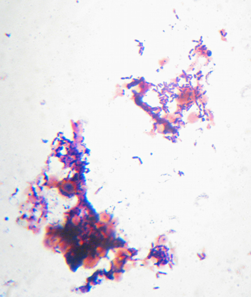
A Gram-stained view of yeasts and bacteria in a sourdough culture named 'Fred'.

I had to do a Fred Transplant last week. A grey fuzzy mold had taken up residence in on the sides of the jar above Fred’s liquid culture, so I set up a fresh container with fresh water and flour, and dipped a spoon down the center of Fred to the bottom, pulling up just a tiny amount of the stuff in there. Then I mixed it into the fresh stuff and covered it with plastic wrap (instead of a paper towel this time.)

Fred smells like Swiss Cheese Feet right now, but he’s obviously still growing, as you can see from last night’s “Gram Stain” microscopy. The slightly blurry light-red-brown lumps are, I believe, yeast cells, possibly Saccharomyces boulardii, since I dumped a capsule of supposedly-still-viable “probiotic” yeast of that species into Fred previously. I have no idea who the bacteria are in here at the moment. I did also see a small number of longer, thinner bacterial cells in there (presumably Lactobacillus) though most of them are the ones you see here.

Meanwhile, I’m about to dig out the still-unused Hillbilly Autoclave and try it out on the media I’m mixing up to try to obtain a culture of genuine wild “native flora” vinegar/kombucha yeast-and-bacteria to play with from the local wildflowers that are just now getting into full bloom.

My starting recipe goes something like this: I mix up about 2 Liters of distilled water with about 100g of glucose (“Dextrose”/”Corn Sugar”), 100g of sucrose, 500mg of L-Arginine, and enough phosphoric acid to drop the pH down to about 5.5 to 6.0. That is intended to be then poured into small “canning” jars in about 100ml amounts and pressure-cooked for at least 15 minutes to sufficiently sterilize and seal them. Meanwhile, a single generic-brand children’s chewable vitamin is crushed up and dumped into a 4-oz bottle of cheap vodka and well shaken.

Then when it comes time to go bioprospecting, I’ll pop open the jar of acidic sugar solution and add about 5ml of the cheap-vitamin-vodka to it to give me about 2% ethanol, and then go find some flowers to cut off and dump into the jars, which will be loosely covered with foil (to let air in but keep dust out) and put in a nice quiet cupboard to grow for a few days.

Hypothetically, the only things that are likely to grow in that will be microorganisms associated with vinegar-making. At some point I’ll also make up a batch of sweet black tea and see if I get a kombucha-like culture going in it, and make up some solid media to try to isolate individual microbes from it.

Microscopy Preview

Intentional food microbiology:
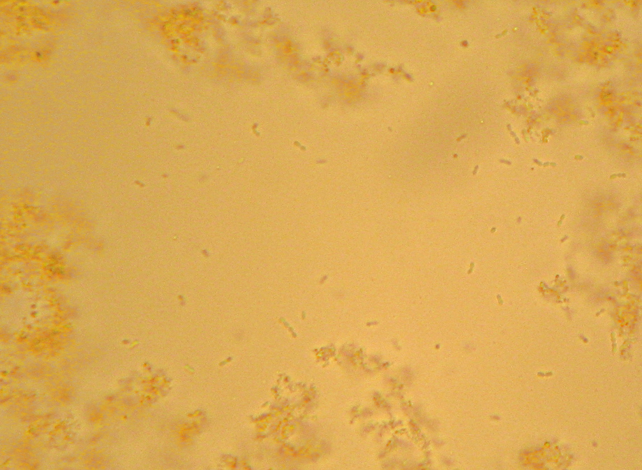
Various yogurt bacteria floating amid milk protein and fat globules

UNintentional food microbiology:
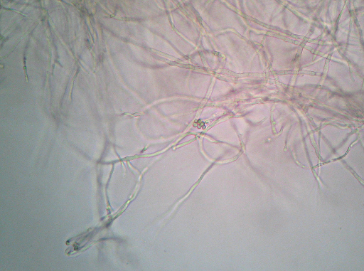
Wet-mount photomicrograph showing mold mycelia and a mass that kind of looks like large spore cases or something

I still don’t feel like I got nearly enough productive stuff done this weekend, but I did manage to do a bit of microscopy – plus demonstrating to myself that I still remember how to do a “Gram stain”. Real Post with explanation and more pictures to follow Real Soon Now…

Amateur Soap Microbiology and my new Friend

I thought soap was supposed to be *clean*!
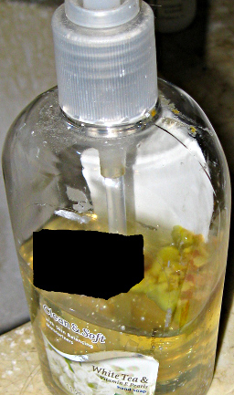
lumpy yellow microbial colonies growing on the soap inside of a hand-soap dispenser
People usually assume soap gets rid of funky microbes that might grow on things, so I was very amused several months ago when I spotted something growing on top of the soap in one of the household hand-soap dispensers. As of today, it looks as pictured at left. That lumpy yellow and brown mass atop the the soap looked to me like some sort of soap-sodden mold, and have been saving the dispenser specifically in the hopes that someday I’d have a microscope and could take a look at it. Meanwhile, the mass spread, and slowly started releasing some kind of yellow pigment into the soap.

Incidentally, I kind of doubt this indicates some sort of failure on the part of the manufacturer of the soap. I don’t recall for certain, but I think I may have opened the dispenser at one point to transfer some of the soap to another nearly-empty dispenser. When the mass started growing originally, it was a single spot, which suggests a single spore or speck of dust floating in and landing on the surface. Hey, it happens. Anyway, I’ve therefore blanked out the name of the manufacturer since I don’t think they really have anything to do with this.
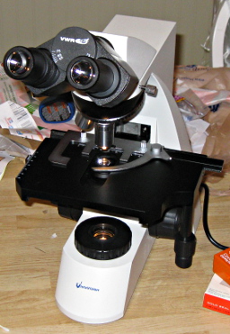
VWR VistaVision Microscope
This mysterious growth upon my soap remained mysterious until today. Thanks to the Minister of Domestic Affairs and VWR (who managed to find me a really good deal), I finally got to actually get a close look at that lumpy mass. Meet my new friend Minnie (pictured at right). I could gaze into those eyes for hours. I couldn’t afford a darkfield condenser, and I sure as heck couldn’t afford to upgrade to phase-contrast gear, but I can add either one later if the opportunity presents itself. I also can’t afford the overpriced proprietary digital camera attachments either, though working around that is a whole other project. Until I identify an affordable model that plays well with Linux or work out how to modify a webcam into an ocular attachment,
I’ll have to settle for a trick…

It turns out if you take a digital camera and set it for close-up photos, you can actually stick the camera lens right up to the eyepiece and often get a serviceable picture.. Now, I had to subject the pictures I got today to moderately heavy processing to bring out the detail a bit better, but at least part of that is just me working on learning how to optimize the camera settings for this kind of use.

Equipped with some surplus slides and cover-slips donated by a kind professor who had some extra packages, I opened up the soap container and smeared a little of the yellow crud on a couple of them. One I just slapped a coverslip on for direct observation – the other I smeared over a slide and let dry with the intention of staining using the tiny, previously-unused vial of methylene blue left over from a very old plastic toy microscope. While the latter dried, I took a look at the wet mount hoping to finally see the mold mycelia that I had been expecting…

There wasn’t enough contrast to bother trying to get a photo, but it was obvious at 400x that what I was looking at was bacteria, not mold. Nerdly joy at learning something by looking in the microscope that I wouldn’t have otherwise known ensued, along with happiness as I realized this meant I had a perfect excuse to dig out my recent shipment from the Maker Shed – materials for doing a “Gram Stain”. Incidentally, the “Maker Shed” had the supplies on the way to me within hours of my ordering it, and they have lots and lots of cool stuff. I highly recommend it. Anyway, I got to do a “Gram Stain” for the first time in a couple of years (and the first time ever outside of a school lab). Want to see?

Mystery Microbe, I see you!
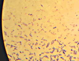
Gram-stained bacteria
Here it is – the nasty yellow goo that infected my bottle of hand-soap. My staining technique was a little off since I’m out of practice – the way I interpret the results is that what I’ve got here is neither a member of the Firmicutes (i.e. “Gram positive”) nor – probably – Actinobacteria. I really can’t guess at more than that, though. I think the few “Gram-positive”-looking cells there are artifacts of insufficient decolorization. I know I still had a surplus of the purple “Crystal Violet” stain still on the slide at the end. (How did I know? I’ll show you at the end…). The irregular bluish bits towards the bottom are, I believe, just bits of stuff from the soap itself.

Meanwhile, this pretty much satisfies my curiousity about the Mystery Soap-Infecting Microbe. There’s certainly a lot more I could investigate, but my developing Hillbilly Biotech lab is really intended to support my interest in intentional food microbiology and perhaps evenutally some small-scale non-food industrial microbiology. I have some remaining curiousity about the yellow pigment and whether or not it might be useful for something, but I’m doubting there is any food or beverage I might want to grow this stuff in and therefore don’t have much use for it. Still, I’ll keep the bottle around for a while before I throw it out in case I think of something fun to do with it. If I end up being really interested in the identity of the bug growing on it, I should be able to find a liquid that I can grow a big mess of it in, then run it through a simple DNA extraction process. Then all I need to do is find someone who can supply PCR primers, a thermocycler, and sequencing services cheap. It might sound like I’m being facetious, but I wouldn’t be surprised these days if I manage to find somewhere that’d do it for $20/sample or less. I may eventually do this will the Mystery Soap Bug anyway, since I hope to be running through this process with cultures of sourdough, yogurt, cheese, vinegar, and brewing microbes that I develop myself. For now, though, it’s just nice to be playing with microbiology equipment again. And now fully independently! Wheeeeeeee!!!!!!

Yes, I’m a nerd. And proud of it!

What’s next?

Now that I finally have a microscope, I no longer have any excuse for not getting to work on the rest of my Hillbilly Biotech lab. Just this weekend I was pricing out Hillbilly Autoclaves. I picked up a cheap air pump and air stone
for potentially building an aerobic bubble-column fermenter (for quick growth of yeast starters or a working model of a “Fring’s Acetator®”-style vinegar generator. I still want to build an ozone generator for sanitization and to get a pH meter. I’d like to also get my hands on some wheat, barley, and rye seeds to sanitize, sprout, and grow here as the first stage of developing a truly local sourdough culture, plus arrange to have several pounds of plain flour irradiated to sterilize it.

I’m also like summer to be over. Yes, I’m writing this in Winter, but it’s not until later in the summer to autumn that locally-grown fruits will start becoming available, and locally grown fruits ought to be an ideal source of local brewing and baking yeasts and bacteria. Finally, I’d like to find a wealthy patron (or matron, I’m no sexist…) who would sponsor me so I could just pursue food-microbe bioprospecting and research full-time…

Oh, yes, and I need to get around to finishing Episode 4 of my little podcast project, especially since episode 4’s topic is a fundamental microbiology technique.

Comments welcome below – thanks for reading!

Oh, and as a reward for getting all the way to the end, here’s a picture that I thought was pretty – crystals of “Crystal Violet” and iodine. I told you I had too much left on the slide…
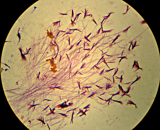
Crystallized dye left on the slide

Ewwwwwwwwwwwwww……

They say “When life gives you lemons, make lemonade”. What if life gives you snot instead?

Someone's slime-covered handNow, see, this is what happens when I’m too poor to buy nice distracting new toys for myself. (No, not the hand in the picture – that’s not mine, it’s just there for illustration.)

I found a pot that I’d rinsed well but then left filled with water in the sink to soak, to help remove the last of the rice bits stuck to it. It hadn’t gotten stinky or fuzzy or anything, but it had gone…viscous. Like a light sewing-machine oil. Naturally, I took appropriate action to deal with it.

I fed it.

Glucose (“dextrose”), to be precise. It’s since been dumped into an old glass jar and the original pot thoroughly scrubbed with hot soapy water. At this point (a day later) the slime is closer to the viscosity of vegetable oil now. And I fed it again.

I wonder what it is? I mean, obviously it’s bacteria-snot, but what kind? I suppose if I had some iodine I could check to see if it’s a polysaccharide (evidently this test works on polysaccharides besides starch). If only I had a microscope, I could at least get some basic hints as to what’s producing the slime. Maybe I can maintain a culture and figure it out later, if I can ever afford a real microscope. Perhaps I could even attempt a strain-improvement program to increase the production rate…

Uh…I did mention I was a nerd, right? Okay then.

I wonder if anyone at work has a bacteriological microscope setup that I could use?…

“Untersuchungen über Bacterien”

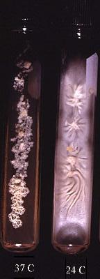
Cultures of Blastomyces dermitiditis, showing how it grows like a mold at one temperature and like a yeast at another.Once again I’m down to the last minute, trying to juggle too many things and almost missing this month’s “Giant’s Shoulders” blog carnival. Almost.

Today we go once again all the way back to the Victorian era, to see that if you thought bacterial taxonomy was difficult now, imagine what it was like over 130 years ago:
Cohn F:”Untersuchungen über Bacterien”; Beiträge zur Biologie der Pflanzen; 1875, vol 1; pp 127-222
(Or “Researches regarding Bacteria”, in “Contributions towards the Biology of Plants”)

This paper is an overview of the problem of categorizing bacteria among the types of living things, and makes some early suggestions. I don’t think it’ll spoil too much of the punchline to point out that not only is the journal about the biology of plants, but the paper also starts out with Cohn describing how he came to work at the “plant physiological institute”. Cohn’s assertion that bacteria are definitely a form of plant actually stuck for at least another three-quarters of a century or so – I have a copy of a 1945 book on bacteriology that actually has a short discussion on why bacteria are categorized as plants rather than animals (or “animalcules”, even). That’s only part of what’s interesting about this paper, though.

Cohn discusses a number of problems with the nature of bacteria in his time. For one thing, he says there had been little real effort to even come up with a coherent scheme for classifying bacteria at that point. He does mention one previous attempt to come up with a system, but on the whole it seems everyone is just coming up with terminology on the fly – even taking Pasteur himself to task for throwing around a variety of terms related to microbes without distinguishing what the terms actually refer to. The reason for this, really, is just that figuring out pretty much anything in detail about bacteria was a seriously difficult problem at the time. Cohn explains why; how it is really impossible to make out more than general shape and size from microscopic examination, and how the lack of any detectable sexual reproduction makes it impossible to do positively identify members of the same species. In fact, even very obvious differences in appearance might not be definitive. It was suspected (and later demonstrated) that some of what appeared to be completely different fungi were actually just different life-stages of the same fungus. (Hopefully you can see the picture at upper right, with the bacteria/yeast-like growth on one tube and the obvious and very different fuzzy mold-type growth on the other. Both are actually the fungus Blastomyces dermitiditis.) Just as some of Cohn’s contemporaries considered that perhaps all molds and yeasts were really just different stages of life of the same organism, perhaps the same might also be true of bacteria?

Cohn does, after all, promote the notion of bacteria as a type of fungus. You may even remember an old word for bacteria: Schizomycetes, that is “fission-fungi” (that is, fungi that reproduce by splitting in half rather than producing spores). This makes sense if you consider that bacteria are more like plants and algae than animals, and fungi were considered to be plants that lacked chlorophyll. Although lamenting that it was not feasible to really separate out individual bacteria to determine whether they ever changed form – this was still three years before Joseph Lister actually did so – Cohn unwaveringly felt that bacteria were in fact made up of several different genera and species, and set out an early attempt at classification.

Once again, Our Friend the American Society for Microbiology hosts a translation of this paper, complete with a couple of paragraphs of more modern editorial commentary at the end. It’s well worth a look.

A photographic portrait of Ferdinand Julius CohnUnfortunately, I don’t think Ferdinand Cohn’s hairstyle is nearly as spiffy as Eduard Buchner’s cool “Colonel Sanders Guest Stars on Miami Vice” look. I think he looks kind of like a slightly-better-fed Sigmund Freud with a bad comb-over. But that’s just me.

“Top Ten Favorite Microbes” proto-meme…

Dr. Joseph over on the “(It’s a…) Micro World (…after all)” blog posted a list of his ten favorite microbes. After showing up in the comments of his post and being a wiseass about E.coli and Gram staining, the least I can do is participate here. Besides, it’s a great idea. Therefore here are ten of my current favorite microbes:

Continue reading “Top Ten Favorite Microbes” proto-meme…

Phylogenetic structure of the prokaryotic domain: the primary kingdoms.

Ike’s comin’ right for us, so I don’t know when the my power and internet access will die, and if so how long it’ll be before it comes back. However, while I’m still connected I wanted to contribute something again to this month’s The Giant’s Shoulders blog carnival. Since it’s in three days and there’s a chance our power might be out when the deadline passes, I figured I’d better hurry. Because of the hurry there are no fancy graphics nor even too much explanatory text here, but I’ll do what I can. Fortunately, the basics of today’s post isn’t too complex.

Depending on how rigorous your biology education was, there are a variety of ways that you might tend to categorize the fundamental types of living things. You might vaguely recall something about “five kingdoms”, which as I recall were “Animals, Plants, Fungi, Protozoa [e.g. amoeba], and Bacteria”. You might just segregate everything into either “animal” or “plant”. If your memory of biology education is a bit stronger, you might remember that “bacteria” are a separate group from the true plants and animals. A step more precise and you may split living things into the two domains of “prokaryote” and “eukaryote”.

The “plant” and “animal” distinction is pretty classic – until comparatively recently, bacteria were assumed to be “plants”, just as fungi (“plants” that lacked chlorophyll) were. Non-photosynthetic bacteria were referred to as “schizomycetes” (literally “fission” [splitting in two] fungi, because they reproduce by splitting from one cell into two rather than forming spores), while bacteria with chlorophyll (cyanobacteria or “blue-green” algae, and possibly the “green sulfur bacteria”) were designated “schizophyta” (“fission plants”).

Within the last fifty years or so, though, it’s become obvious that bacteria were a different type of life from fungi, chlorophyll-containing plants, or animals. The latter critters have cells that in turn contain “organelles”, which are more or less very specialized “mini-cells” within themselves. The nucleus, for example, is a compartment within the cell where the cell’s DNA is kept and processed. Bacteria, it turned out, don’t have any of these organelles (in fact there’s good evidence that at least some if not all organelles used to be bacteria, but this post’s long enough already so I won’t go into that), and life was re-organized into the bacterial “prokaryotes” (“before nucleus”) and the “eukaryotes” (having a “true nucleus” – i.e. everything that isn’t bacteria).

Then, along comes Carl Woese, who spoils this nice simple dichotomy. In 1977, he published (along with G.E. Fox) the subject of today’s post:
Continue reading Phylogenetic structure of the prokaryotic domain: the primary kingdoms.

“Ueber die isolirte Faerbung der Schizomyceten in Schnitt- und Trockenpraeparaten”

The Giant’s Shoulders blog carnival is coming up in two days, and I just realized I still haven’t gotten a post up for it yet. So, here it is.

I put up some quick reviews of several classic microbiology-methods papers for the previous edition of this blog carnival, but didn’t actually get around to putting up the one for what is almost certainly the most well-known microbiology technique: “The Gram Stain”. So, this post is about it:

Gram HC: “Ueber die isolirte Faerbung der Schizomyceten in Schnitt- und Trockenpraeparaten”; Fortschritte der Medicin; 1884; vol 2, pp 185-189

That’s “Regarding the Isolational(?) Coloring of Schizomycetes in Cut- [i.e. tissue sections] and Dried Preparations” in “Medical Progress”. The translation hosted by the American Society for Microbiology uses the word “Differential” where I’ve put “Isolational” – which is probably not quite right either but it’ll have to do for now – but I’ll get to that in a moment.

If you’ve ever been exposed to microbiology labwork before, you’ve almost certainly done or at least watched a procedure referred to as a “Gram stain”. In brief, you smear your sample with bacteria on a glass slide and bake it on, then you dump some purple stuff on it, them some brown stuff, then you rinse it briefly with alcohol, then you dump on some pink stuff, and then rinse it in water and look at it under a microscope. Bacteria that stay the original dark purple-blue color of the original purple/brown stuff are considered “Gram Positive”, and those that don’t instead appear the pink color of the last stain, and are considered “Gram Negative”. Many textbook authors and microbiology instructors will breathlessly proclaim that the Gram Stain reveals two “fundamental” categories of bacteria, but I’ll spare you my rant about that.

Properly speaking, this isn’t actually Gram’s stain, as described in his original paper. The modern variations that we’re all taught in microbiology class were developed later, and I believe they are nowadays based mainly on Victor Burke’s 1922 paper on the subject[1].

Regarding the title of the paper: “schizomycete” is what they used to call most kinds of bacteria. “Mycete” meaning “fungus”, as bacteria were assumed to be “plants without chlorophyll” just like molds and mushrooms, and “Schizo-” meaning “split in two”, since bacteria reproduce by splitting into two cells rather than by producing spores like “other” fungi. I say “most” because things like cyanobacteria (“blue-green algae”) or Green Sulfur Bacteria would have been referred to as “Schizophyta” (“fission-plants”). What Gram was originally trying to do wasn’t to differentiate one kind of bacteria from another, either, but to make it easy to tell bacteria from from the nuclei of cells in bacteria-infected tissue.

For that matter, Gram was really metaphorically standing on the shoulders of Koch and Erhlich, as he was building on their technique for staining “tubercle bacteria” – that is, tuberculosis-causing members of the genus Mycobacterium. Gram mentions that you need to stain this type of bacteria for the “usual” 12-24 hours to make this work, incidentally, as opposed to a few minutes for other “schizomycetes”. This suggests that you are expected to have some idea of what you’re going to find before you use the stain, as opposed to the modern implementation which is supposed to tell you something about what kind of bacteria you’re finding.

Still, Gram does report that some bacteria take the stain and some don’t, giving us a preview of the “differential” character of the modern version. He specifically notes typhoid and some causes of bronchial pneumonia fail to hold the stain. Given that Typhoid Fever is caused by a strain of the “Gram-negative” butt-bacter Salmonella enterica, and there are a number of “Gram negative” bacteria as well as “Gram positive” that can cause pneumonia, this makes sense. He also does mention the use of Bismarck Brown R a.k.a. Vesuvine as a counterstain in order to make the nuclei of the infected cells brown in contrast to the dark blue of the infectious bacteria in the tissue.

For much of the century-and-a-quarter since Gram’s publication, the question of why the Gram stain works was thoroughly investigated, and even today I occasionally hear or read assertions to the effect that the Gram Stain isn’t well understood. I disagree with this just as I think its importance to bacterial identification is grossly overblown, and if you want to know why, I have a previous post all about why the Gram stain works and how we know. You may or may not also be interested in an older post regarding whether or not “acid-fast” bacteria like the ones that cause tuberculosis (which don’t stain at all when using the modern version of the Gram stain) are “Gram Positive” or not. As always, if you spot any errors or have any questions, please let me know…

[1] Burke V: “Notes on the Gram Stain with Description of a New Method.” J Bacteriol. 1922 Mar;7(2):159-82.

Benzoic Acid Part 2: “Sour Stuff”

Okay, now that the boring review is over with…

Consider the cell. It doesn’t matter what kind of cell – bacterial, archael, fungal, animal, whatever. It’s still a tiny droplet of slightly salty water, thickened by a bunch of enzymes, other proteins, and various other substances floating around in the water. There’s also one other component that makes this a “cell” rather than soup: a bubble made of fatty material that the droplet is wrapped in, called the cell membrane. Depending on what kind of cell you’re thinking of, there may or may not be a “cell wall” made of some sort of rigid material, with the cell membrane inside of it. There may also be more than one membrane as is the case with the classic “Gram negative” style of bacterium, which has a second “outer” membrane wrapped around its cell wall. If it’s a eukaryotic cell, it’ll even have tiny little “organelles” inside itself wrapped in their own little membranes…but whatever. It’s the innermost one, inside of whatever cell wall may be there but wrapped around the cell’s guts, that we’re concerned with here.

Since stuff that will dissolve readily in water doesn’t tend to dissolve well into fats, and vice-versa, the cell membrane not only prevents stuff dissolved in the water inside the cell from leaking out, it also prevents stuff in the water outside from getting in. This lets a cell maintain itself at near neutral pH even if it happens to live in a very acidic environment, or an appropriate level of, say, sodium salts even if it lives in the Great Salt Lake.

This brings us back again to benzoic acid, which you should recall from the previous post alternates between a dissociated hydrogen-ion-and-benzoate-ion form and a combined, netural form in water. You may have noticed that foods preserved with benzoates tend to be sour, like fruit juices or soda. That’s because “sour” is the flavor of acid, and benzoic acid’s ability to be a preservative is only good in acidic environments

Useless Knowledge Break: the German word for acid is “Saurstoff”. Yes, that is pronounced like “sour stuff”, and no, that is not a coincidence.

An acidic environment means lots of extra hydrogen ions (“protons”) floating around. That also means that when a molecule of benzoic acid splits into a hydrogen ion and benzoate ion, it takes less time before another hydrogen ion comes by and the molecule can recombine again and therefore a bigger majority of the benzoate floating around at any moment is in the combined, somewhat fat-soluble neutral form. In that form, it can soak into a cell membrane if it encounters one.

If that molecule drifts through the membrane and gets to the inside of the cell, it may touch the less acidic watery environment there and dissociate into ions again and be unable to return through the membrane. The released hydrogen ions mean the inside of the cell becomes more acidic. As of today (20080806), the Wikipedia entry for Sodium Benzoate cites a single paper from the early 1980’s saying that when the inside of a yeast cell gets acidic enough, it prevents a specific step in the energy-generating process from working. This may be true, but there’s more to the story than this.

Obviously the membrane can’t totally seal the cell off from the outside, or the cell would be unable to excrete wastes or take in food molecules, so there are numerous specialized “transport” proteins that stick through the membrane to allow specific kinds of molecules in and out. Lots of biochemical reactions release hydrogen ions, so there are transport proteins that can shove hydrogen ions out of the cell and into the cell’s surroundings. The problem is that all substances naturally diffuse from areas of higher concentration to areas of lower concentration, so in an acidic environment the natural direction that hydrogen ions “want” to flow is into the more neutral cell. These transport proteins can shove the hydrogen ions in the opposite direction, but like pushing a boulder uphill it costs energy. This seems to be the primary reason that benzoic acid prevents bacteria and yeasts from growing – it makes them waste energy that they would be using for growth just to keep taking the hydrogen ions that the benzoic acid helps leak in through the cell membrane and shoving them back outside. The figure above is linked to a page at Helsinki university that discusses this type of preservative action in more detail.

Simple and elegant, and this seems to have been assumed to be the whole explanation for some time. But what happens to the benzoate ion when its hydrogen ion gets pumped away? Does it do anything?

Coming up next: Endocannibalism!

“A small modification of Koch’s plating method.”

Only two more days for the Classic Papers Challenge, so if I’m going to get any more up, I’d better get my butt in gear.

Here’s a nice easy one:

Petri, R. J.:”Eine kleine Modification des Koch’schen Plattenverfahrens.” Centralblatt für Bacteriologie und Parasitenkunde; 1887; Vol. 1, pages 279-280.

The American Society for Microbiology has a translation available online. It’s only about a page-and-a-half of relatively large type – check it out.

There’s a trick we microbiologists use for growing bacteria. You make a solid (but wet) surface that contains whatever nutrients the microbe (bacteria, archaea, yeasts, mold spores…) you’re interested in need, and then you spread a diluted mixture of the microbe on it. The idea is that since the surface is solid the microbes can’t move around too much, and at any spot where a single cell starts initially, a whole pile of that cell and it’s genetically-identical (non-sexually-produced) clone-children will form until it gets big enough to see without a microscope. This cell-pile is called a “colony”, and you can poke or rub it with a sterile object, then stick the object into a sterile nutrient source. The end result is you have a “pure” culture of microbes that are effectively genetically identical. The solid material could be a lot of things – I’ve seen references to using slices of potato – though these days agar-agar gel mixed with nutrients is the preferred substance.

Koch (that is, Robert Koch of “Koch’s Postulates” fame, not Ed Koch the former mayor of New York City) used gelatin (so, hey, here’s another thing you can do with your expired Jell-O®). He apparently used to have a stack of shallow bowls, and had to use a special pouring device to carefully dump the gelatin into each stacked bowl in turn, then cover the works with a bell jar in order to keep stuff from falling into them from the air and contaminating them.

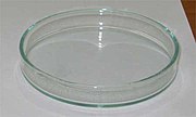
This was kind of a pain to work with, so some clever guy named Julius came up with a modification of this method in 1887, using pairs of shallow dishes, one slightly larger than the other so that it could be turned upside down to use as a lid. Then, you don’t necessarily need the bell jar, and you don’t need to stack them so they’re easier to pour.

Julius Robert Petri’s idea was so useful that we still use it today. Oh, yeah, and they named the dish-and-lid combination after him.

How’s that for a “classic” paper?

Meanwhile, my “Mountain Dew® Wine” project is turning out to be substantially more educational and fascinating than I’d hoped. There seems to be a decent amount of information available on how benzoic acid affects yeasts. I intend to turn that into a post later, but first I’ll try to find at least one more old paper to post before tomorrow is over…